Cell Analysis & Imaging Lab: 9 Resources found
| CAI Technical Assistance | Industry Rates
|
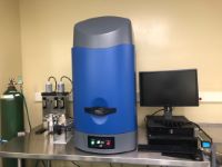
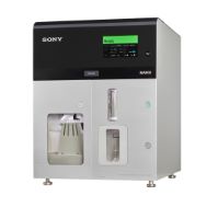 | Cell Sorter Sony MA900 MINIMUM OF ONE HOUR RESERVATION
The MA900 supports a wide range of applications. The system offers flexibility with a choice of 4 excitation lasers - 488 nm, 405 nm, 561 nm and 638 nm on two beam spots. Free-form PMTs enable detection of fluorescence signals from each beam spot, allowing detection of up to 12 fluorescence parameters and 2 scatter parameters. Microfluidics sorting chips are available in 3 sizes 70um, 100um and 130um for sorting into 2-way and 4-way tubes and 96 and 384 well plates. | Industry Rates
|
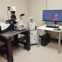 | Confocal Laser Scanning Microscope Leica SP8 Additional pricing for academic and government groups is available upon request.
| Industry Rates
|
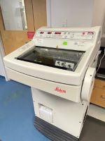 | Cryostat Leica CM1850 Frozen tissue section prep. Self users provide their own slides, slide box, and OCT-embedded tissue blocks. An additional fee will apply to use the slides at the Cell Analysis and Imaging Lab.
| Industry Rates
|
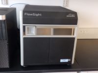 | Flow Cytometer EMDMillipore The Amnis® FlowSight® Imaging Flow Cytometer has three excitation lasers (405, 488, 642nm) and twelve standard detection channels that simultaneously produce brightfield, darkfield or side scatter, and up to ten channels of fluorescence imagery of every event at ~20X magnification with high sensitivity CCD cameras. The FlowSight® provides users with the ability to gain detailed images of a large number of cells in a relatively short period of time and the opportunity to perform a range of novel applications including co-localization, internalization, stem cell differentiation, cell cycle, phagocytosis, and cell-cell interactions.
The instrument configuration includes with fluorescence amplification and minimal noise detection that results in very high signal sensitivity paired with a powerful quantitative image (QI) processing option. Data files can be analyzed using Amnis® IDEAS® software for image processing alongside traditional gating strategies or saved in .fcs format for analysis through alternate software types (i.e. FlowJo or ModFit™). In addition to being able to see the cells displayed in dot plots and histograms, the imaging provides analysis tools not available in conventional cytometers such as texture and spot counting.
Booking the Instrument:
The time required for each sample is dependent on the cell concentration, the number of events needed and the complexity of the compensation strategy. A good general guideline for booking is 10 minutes per sample to start. Additionally, if your experiment requires a DNA dye or potential contaminants to subsequent experiments there will need to be a sterilization step (45min) which needs be included in the booking of the machine by the user. (This will be determined by CAI Staff when Data Acquisition Form is submitted and based on the experimental design.)
We do not currently accept MRSA or BSL-2 samples. | Industry Rates
|
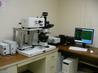 | Fluorescence Microscope 4 Documents AvailableOlympus LSCM FV300 Sample Characterization Equipment
Laser Scanning Confocal
Additional pricing for academic and government groups is available upon request.
| Industry Rates
|
 | IVIS AMI-HT Spectral Instruments Imaging, Inc. The Spectral AMI-HT provides unrivaled sensitivity for bioluminescence and fluorescence in vivo imaging. AMI-HT acquires quantitative images from a diverse set of applications including well plates, plants and small animals. Designed around the needs of animal scientists, the Spectral AMI IVIS Imaging system include a robust build, patented LED illumination source, custom filter options, -90°C cooled camera, and absolute calibration to enable high throughput imaging.
· 5 Mouse Capacity (BLI/FLI) - 25cm x 17cm field of view.
· LED Fluorescence Illumination
· 10 Wavelengths/10 Filters. Additional filters can be added/changed by
operator, if needed
· Mouse-friendly heated Platform allows you to easily recover wandering mice
· Anesthesia compatible with all third party gas anesthesia systems
· -90°C Cooled Camera. Ultra cold for maximum sensitivity
· Absolute Calibration ensures quantifiable data
http://spectralinvivo.com/imaging-systems/
| Industry Rates
|
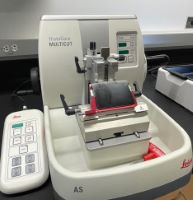 | Microtome Leica HistoCore Multicut Section tissue that has been fixed and embedded in either paraffin or resin. The microtome will enable to cut sections 0.5um to 100um in thickness. Self-users provide their own slides, slide box, and paraffin-embedded tissue blocks. An additional fee will apply to use the slides at Cell Analysis and Imaging Lab.
| Industry Rates
|
 | Paraffin Embedding Console Sakura Tissue-Tek SEC 6 Tissue-Tek SEC 6 is a tissue embedding station which is used to embed tissue samples in paraffin wax. This station features a digital programmable interface which enables to program individual temperature settings for the paraffin reservoir, cassette bath, mold warmer and work surface.
| Industry Rates
|