Microscopy Core Lab: 28 Resources found
 | 2GB Flash Drive Please see MCL staff to pick up this item. | Industry Rates
|
 | Atomic Force Microscope PARK XE-100 Sample Characterization Equipment
Location: Olney OG-3
Training Estimates: 6 Hours
An Atomic Force Microscope (AFM) consists of a Si or Si3N4 microscale cantilever that has a very sharp tip (usually 5-15nm radius). The tip is brought to very close proximity to the sample surface and scanned laterally across the surface. Forces (such as mechanical contact, Van der Waals, capillary, chemical bonding, electrostatic, magnetic, etc.) between the tip and the atoms on the sample surface lead to vertical deflections of the cantilever according to Hooke’s Law. By monitoring the deflection using a laser spot reflected from the top of the cantilever onto a position sensitive photodetector array, one can construct a 3-dimensional topographic map of the sample surface. Typical image lateral resolution is in the order of the tip radius, although atomic resolution can be achieved in non-contact mode, as well. The vertical resolution can be in the order of angstroms. AFM is applicable to both electrically conductive and insulating samples and requires very little sample preparation procedures.
Include Sample Description and Analysis Requirements in note section of Order Form
Additional pricing for academic and government groups is available upon request.
| Industry Rates
|
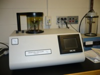
 | Critical Point Dryer Tousimis SAMDRI-795 Sample Prep Equipment
Additional pricing for academic and government groups is available upon request.
| Industry Rates
|
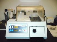
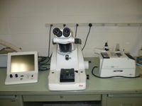 | Cryo-Ultramicrotome Leica EM UC6-FC6 Sample prep instrument for SEM, TEM and AFM imaging
Additional pricing for academic and government groups is available upon request.
| Industry Rates
|
 | Desk IV Sputter and Desk II Carbon Coaters Denton Vacuum Desk IV (Sputter Coater), Vacuum Desk II (Carbon Coater) For SEM preparation by sputtering (gold or other noble metals) and by carbon evaporation.
Additional pricing for academic and government groups is available upon request.
| Industry Rates
|
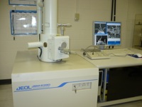
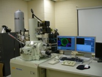 | Field-emission Scanning Electron Microscope 5 Documents AvailableJEOL JSM 7401F Unit 1 Sample Characterization Equipment
Location: Olney OG-1
Training Estimates and Prerequisite:
6 Hours and must have demonstrated proficiency in using JEOL 6390 SEM
Features:
1. Electron optical column: Cold-cathode tip field emission gun; 1.0 nm resolution at 15 kV accelerating voltage; 0.1 – 30 kV accelerating voltage range; 25x – 1,000,000x magnification range
2. Detectors: one chamber-mounted Everhart-Thornley type secondary electron detector, one semi-in-lens secondary electron detector with r-filter and secondary electron signal enhancer, one pneumatically retractable solid state back-scattered electron detector for topographical and compositional image contrast
3. Specimen chamber and stage: large specimen exchange port accommodating 4 inch diameter and 40 mm height samples, eucentric goniometer stage with PC-automated X-, Y- and R- axes and manual Z- and Tilt- axes
4. X-ray Micro-analysis using Energy Dispersive Spectroscopy: EDAX Genesis XM2 Imaging System composed of a 10 mm2 Si(Li) detector with SUTW window for detection of all elements down to Be, and the digital electronics and software for image acquisition and x-ray signal mapping and qualitative and quantitative analysis capabilities
5. Nanometer Patterning Generation System for e-beam lithography includes NPGS PCI 516A high speed lithography board, Deben PCD beam blanking system, NPGS v9.0 control software and DesignCAD LT2000
Include Sample Description and Analysis Requirements in note section of Order Form
Additional pricing for academic and government groups is available upon request.
| Industry Rates
|
 | Field-emission Scanning Electron Microscope 5 Documents AvailableJEOL JSM 7401F Unit 2 JSM 7401F Unit 2 - Manufacturer: Jeol
Saab ETIC building (Room 154 A)
Training Requirements and Estimates: 6 hours and must have demonstrated proficiency in using the JEOL 6390 SEM
Sample Characterization Equipment Specifications:
Electron optical column: Cold-cathode tip field emission gun; 1.0 nm resolution at 15 kV accelerating voltage; 0.1 – 30 kV accelerating voltage range; 25x – 1,000,000x magnification range. Detectors: one chamber-mounted Everhart-Thornley type secondary electron detector, one semi-in-lens secondary electron detector with r-filter and secondary electron signal enhancer, one semi-in-lens solid state backscattered electron detector, one pneumatically retractable solid state back-scattered electron detector for topographical and compositional image contrast
Specimen chamber and stage: large specimen exchange port accommodating 4 inch diameter and 40 mm height samples, eucentric goniometer stage with fully PC-automated X-, Y-, Z-, tilt and R- axes
Energy Dispersive Spectroscopy (EDS), using Oxford Ultim Max Detector composed of a 170 mm2 SSD detector for detection of all elements down to Li, and the digital Extreme electronics and AZtec software for image acquisition, x-ray signal mapping and qualitative and quantitative analysis capabilities
For SEM lab service email Miguel_Matos@uml.edu. Include Sample Description and Analysis Requirements in email.
Additional pricing for academic and government groups is available upon request.
| Industry Rates
|
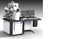 | Focused Ion Beam-Scanning Electron Microscope Zeiss Auriga 40 FIB-SEM Auriga FIB-SEM - Manufacturer: Zeiss
Core Research Facilities Characterization Suite (Room 154 B) - Faraday shield room
Training Requirements and Estimates: 6 hours and must have demonstrated proficiency in using the JEOL 7401F FE-SEM
Sample Characterization Equipment Specifications:
Ga liquid metal ion source FIB specs: 7 nm @ 30 kV, 600x - 500kx magnification, 1 pA - 20 nA for fast and precise sample modification
GEMINI® FE-SEM specs: 1.1 nm @ 20 kV, 20x - 900kx magnification, Thermal field emission type 0.1 - 30 kV, 2.5 nm @ 1 kV for Ultra high resolution and contrast for best SEM imaging, standard and non-conductive.
In-lens EsB® detector and STEM detector
CrossBeam® operation (milling and polishing with live SEM imaging capability completely independently from each other)
Multi-channel gas injection system for selective etching, enhanced etching, material deposition, insulator deposition
Super eucentric, fully motorized stage and dry pumping system
Additional future capabilities:
Omniprobe nano manipulator for the lift-out technique for TEM-STEM imaging
Equip with Leica cryogenic stage for FIB milling a specimen at cryogenic temperatures. Particularly useful for the milling of biological specimens, gels, or low Tgpolymers and allows studies of wet samples with the scanning electron microscope under high vacuum conditions
Note: To become a self user will require training on Zeiss Auriga SEM and FIB features. User must demonstrate proficiency on SEM before continue with FIB training.
For FIB/SEM lab service email Miguel_Matos@uml.edu. Include Sample Description and Analysis Requirements in email.
Additional pricing for academic and government groups is available upon request.
| Industry Rates
|
| General Lab Training Level Two Lab Training
Must be completed prior to use of instruments | Industry Rates
|
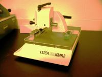 | Glass cutter Leica EM KMR2 Sample Preparation Equipment
no additional charge | Industry Rates
|
| Glow Discharge PELCO easiGlow | Industry Rates
|
 | Grinder-Polisher Buehler Ecomet 250 Ecomet 250 - Manufacturer: Buehler
Core Research Facilities Characterization Suite (Room 154 C)
Training Requirements Estimates: 1 hour
Simple Operation
Touch screen Pro version or tactile feedback membrane control
Easy to actuate blue flashing iridescent Power standby button
Large, red and easily accessible emergency-stop button for safety
Variable Speed Power Head
Single & Central force operation
Variable speed for complimentary or contra rotation
Accessible Work-space
D-style bowl for easy platen changing
Removable splash guard minimizes splashing
Bowl cover protects platen while not in use
Clean-ability
Retractable water hose for wash down
Disposable bowl liner for quick cleaning
Integrated 360 deg bowl rinse
Additional pricing for academic and government groups is available upon request.
| Industry Rates
|
 | Grinding and Polishing System Struers DP-U4 Sample Preparation Equipment
Additional pricing for academic and government groups is available upon request.
| Industry Rates
|
| Lab Staff Additional Costs Technical expertise beyond "Assisted Use" | Industry Rates
|
| Liquid Nitrogen Fill Service | Industry Rates
|
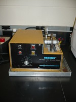 | Low Speed Saw Isomet 11-1180 Sample Preparation Equipment
Additional pricing for academic and government groups is available upon request.
| Industry Rates
|
| MCL Technologist | Industry Rates
|
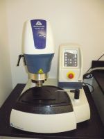
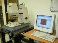 | Optical Profiling system Wyko NT2000
Sample Characterization Equipment
Training Estimates : 4 Hours
Surface heights measurement
1. Phase-shifting interferometry (PSI) mode allows measuring fairly smooth and continuous surfaces (0.1 nm < heights < 160 nm).
2. Vertical scanning interferometry (VSI) mode measurse rough surfaces and heights ranging between 160 nm and 2 mm.
Include Sample Description and Analysis Requirements in note section of Order Form
Additional pricing for academic and government groups is available upon request.
| Industry Rates
|
 | Precision Saw Buehler Isomet 1000 Isomet 1000 - Manufacturer: Buehler
Core Research Facilities Characterization Suite (Room 154 C)
Training Requirements Estimates: 1 hour
A wide selection of chucks, fixtures, and rotating vises guarantee proper fixture
Gravity feed design allows for reproducible and minimal sample deformation
Can be set to turn off at a predetermined cutting depth
Optional rotating chuck decreases heat from cutting
Table saw attachment enables manual sectioning of larger parts
Removable coolant tray for fast cleaning and easy removal of cut samples
Blade dressing device available for rapid dressing during cutting
Acceptable blades: 4in, 5in, 6in, and 7in diameter
Additional pricing for academic and government groups is available upon request.
| Industry Rates
|
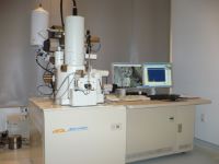
 | Scanning Electron Microscope JEOL JSM 6390 Sample Characterization Equipment
Location: Olney OG-2
Training Estimate: 4 hours
Features:
1. Electron optical column: Conventional tungsten thermionic emission gun; 3.0 nm resolution at 30 kV accelerating voltage; 0.5 – 30 kV accelerating voltage range; 5x – 300,000x magnification range
2. Detectors: one chamber-mounted Everhart-Thornley type secondary electron detector for both SE and BE imaging
3. Specimen chamber and stage: large specimen chamber accommodating up to 6 inch diameter wafers, eucentric goniometer stage with manual X-, Y-, Z-, R- and Tilt- axes
4. Automated features: Auto Focus/Auto Stigmation; Auto Contrast and Brightness; Auto gun saturation, bias and alignment
Include Sample Description and Analysis Requirements in note section of Order Form
Additional pricing for academic and government groups is available upon request.
| Industry Rates
|